
- Why Understanding the Brain Is Going to Be a New Renaissance: "Let's Understand Who We Are Inside"
- Neuroscience The first 'Brain Atlas' to understand neurons cell by cell has been set up
The publication, a few months ago, of the first atlas of the human brain was a very important step towards understanding how it works and what are the keys to this organ that coordinates the body and houses our mind.
Experts such as Rafael Yuste, professor of Neurobiology and Neuroscience at Columbia University (USA) predicted the arrival of "a new Renaissance", a revolution in neuroscience that would allow us to build, in the medium term, a general theory about the structure, connections and function of the brain.
"We've been gutting the molecules and cells in the brain for 100 years, but what we haven't done yet is put all the pieces of that puzzle together. However, we are beginning to see light at the end of the tunnel, there is beginning to be a general theory of how all these neurons fit together and what the brain is for," Yuste said in this recent special on the brain published in EL MUNDO.
"It's similar to what happened with genetics. Scientists had also been accumulating data for about 100 years, putting all the pieces of the puzzle on the table, but they didn't know how to fit it together until Watson and Crick, actually also with Rosalind Franklin's stolen data, came the model of the double helix of DNA. I think a similar thing is going to happen in neuroscience. There will be a general theory that brings together all this data that is now floating around and that will allow scientists and doctors not only to understand how the brain generates the human mind, but to understand brain diseases and to be able to tackle and cure them," he said.
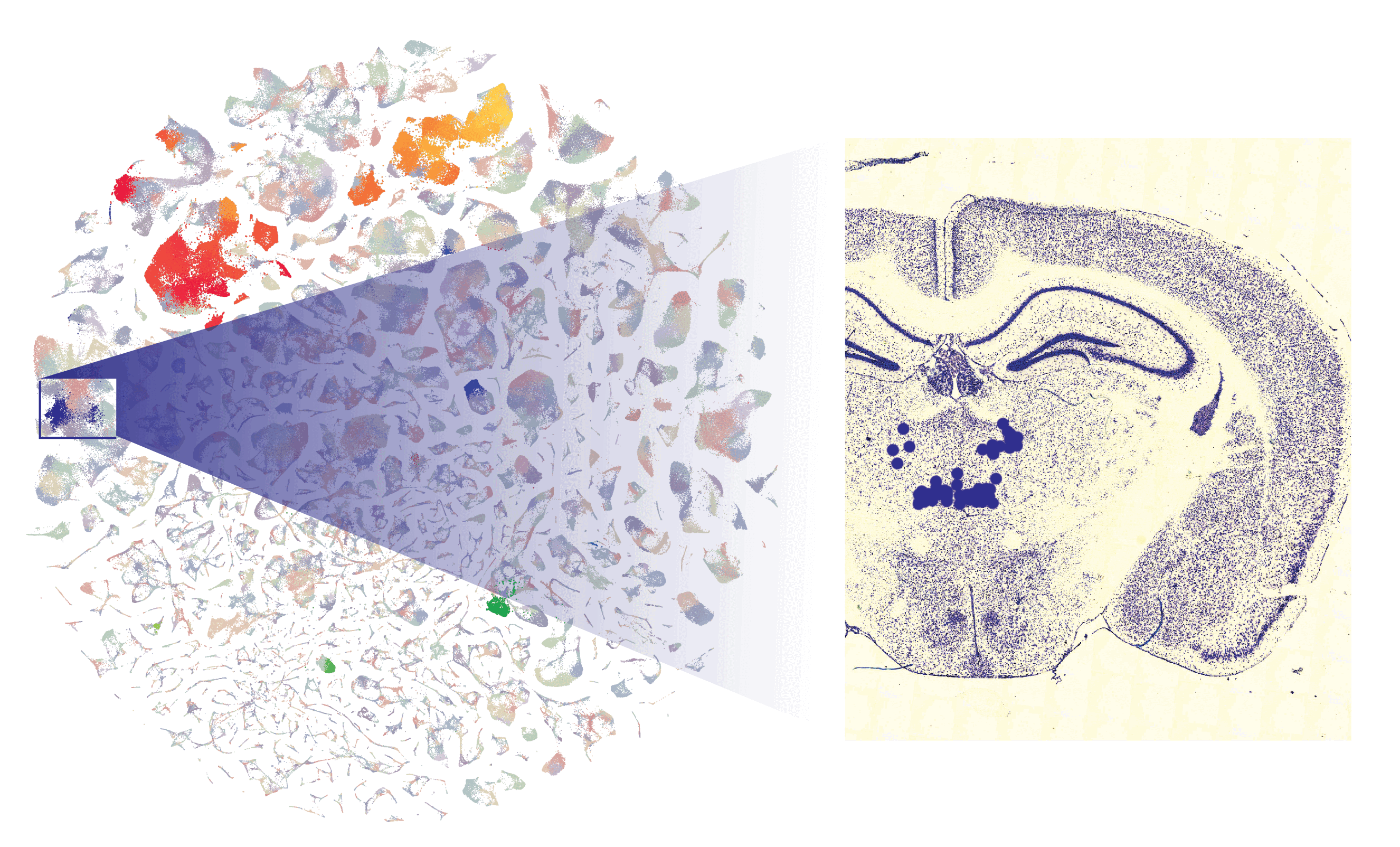
That pathway to a better understanding of the brain expands this week with the publication in the journal Nature of nine studies that, together, provide the most complete and detailed characterization of the mouse brain.
Why is it important to have the mouse brain map?
This mapping provides fundamental data on the structure and organization of the brains of these animals, as well as on the functioning of neural circuits and the function of brain cells at the individual level.
These new data will serve as a tool, as the scientific journal emphasizes, to advance research on the development and evolution of the mammalian brain, which has implications for understanding the genesis of neurological disorders.
"Echoing previous articles from just a few months ago, this batch of articles is history again. It is another of the first salvos of what will be a torrent of studies in the next decade that will classify, using transcriptomics techniques, all the cell types of the body," Yuste told Science Media Centre Spain in the wake of the Nature publication.
For the specialist, the most innovative thing about the new contributions is that "not only are the cell types of the brain mapped, but the position of each type of neuron is mapped for the first time. This is what is called spatial transcriptomics, using new microscopy techniques that allow transcriptomic analysis of certain positions in histological sections."
"These data are not the end but the beginning of the road," warns the researcher, who then points out the limitations of the new studies: "the information is only from the soma [body] of the neurons, it is a still photo of the genes that were active at a given time and they do not tell us which ones were important. Much work remains to be done. In any case,this consignment of items is impressive," he concludes.
And how does knowing the brain of a worm help us?
Another paper recently signed by researchers at the Laboratory of Molecular Biology of the Medical Research Council in Cambridge (UK) also represents an important advance in the field of neurobiology. These scientists, including the Spaniard Lidia Ripoll, have managed to reveal the first wireless map of a worm's nervous system.
Specifically, they have managed to detail how communication occurs in individuals of C. elegans through neuropeptides, small proteins with action on the nervous system. Oxytocin or thyroid hormone are some of these molecules that are capable of generating connections between neurons that are not next to each other. Your networks, therefore, can be thought of as a wireless connectome.
Ripoll uses an analogy to explain this: "Let's imagine that neurons are towns or cities. The synapses would be the highways, the standard paths that connect these towns and cities to each other. But in addition to these roads, there are other ways of communication. Neuromodulators act as traffic signals, such as traffic lights or speed signs that allow connections to be created between towns and cities that are not connected by a road," he explains. In addition, these signals can act as switches, for example by increasing or reducing the strength of a synapse, adds the researcher.
Understanding how these wireless networks work can be key to understanding issues such as the neural basis of behavior or finding drug targets for different disorders.
"The basic mechanisms of neuropeptide signaling are shared by all animals: neuropeptides are released from neurons and diffuse to distant areas to bind to receptors on other neurons," says Ripoll, who remarks that "the nervous system of the worm is anatomically small, but at the molecular level its neuropeptide systems are very complex. showing significant parallels with those of larger animals. We hope that the neuropeptide connectome from C. elegans will serve as a prototype for understanding wireless signaling in larger nervous systems."