
— Your research group published a scientific article in the Journal of Imaging, which is dedicated to a computer program that you developed to analyze data on the activity of neurons in the brain. Please tell us more about it.
"Miniature microscopy allows us to collect a large amount of data on the activity of neurons and neural networks. Therefore, scientists are faced with the urgent issue of processing and analyzing these arrays of information. Most recently, we published a scientific article in the Journal of Imaging, in which we presented to the scientific community the NeuroActivityToolkit program we developed. It allows you to quantify the data obtained with the help of a miniscope. NeuroActivityToolkit is a set of tools for statistical processing of neural network activity indicators. The program is freely available, and other researchers can use it to process their experimental data. The software allows you to assess changes in the activity of neural networks under the influence of age, external influences or pathologies. The latter is especially important to us as our research team conducts research related to Alzheimer's disease.
- Evgeny Gerasimov
- © SPbPU Press Service
What are neurons, how do they combine into neural circuits in the human or animal brain, and how do they function?
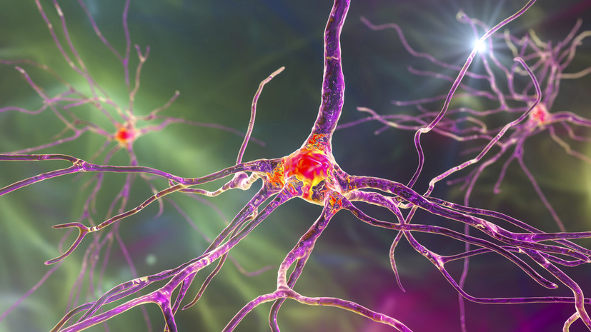
Neurons are excitable cells of the nervous system that transmit information using electrical impulses. In the brain, connections between neurons are carried out through nerve endings called synapses. On one side of each neuron there is a special process, the axon, which sends a signal, and on the other side, a dendrite is a process that receives signals. The dendrite has special protrusions of the cell membrane called dendritic spines, which are able to collect and integrate signals from other nerve cells.
Signals are divided into excitatory and inhibitory, and together their balance ensures the stable functioning of the brain and the functional representation of its different areas. Also, the functioning of neurons is supported by glial cells, which are auxiliary brain tissue. It includes several types of cells, such as astrocytes and microglia. For example, it is known that astrocytes are able to influence the transmission of nerve impulses by neurons, and microglia are involved in the brain's immune response.
Each neuron is connected to a large number of other cells, which together form a complex neural network. In this way, intricately organized networks of interconnected neurons that control most high-level nerve functions can be found throughout the brain.
— At the very beginning, you mentioned miniature microscopy. What is this technique and what other ways are there to record the activity of living neurons in the brain?
— To begin with, the most informative research that allows us to understand how neurons work and interact is tracking the neurons of a living animal organism in normal conditions.
- Gettyimages.ru
- © PIXOLOGICSTUDIO/SCIENCE PHOTO LIBRARY
Until recently, there were mainly two methods in the arsenal of scientists that allowed them to observe neurons in real time. Firstly, it is a method of two-photon microscopy, which makes it possible to detect changes in the fluorescence of calcium tracers resulting from the generation of an electrical impulse. The two-photon microscopy method provides high-resolution images, but it has a drawback: in order to count the activity of neurons, the animal's head must be fixed in one position. To simulate the real activity of the animal, it is immersed in a virtual space, where it must undergo various behavioral tests. At this time, the activity of neurons is recorded.
The second method is to record activity using electrode arrays that are implanted in the animal's brain to study the electrical activity of cells. The method is highly sensitive to changes in the action potential of nerve cells. Let me remind you that the action potential of neurons refers to changes in the ionic permeability of the cell membrane, which generates an electrical signal, so that information is transmitted from neuron to neuron.
- Lab mouse with miniscope sensor on head
Miniature fluorescence microscopy (miniscope) can partially overcome these drawbacks. The sensor of the device weighs only three grams, which allows you to attach it to the head of a laboratory mouse, while you do not need to fix the animal. The resolution of the device makes it possible to visualize individual neurons and record changes in their activity using genetically encoded calcium tracers. These are special proteins that are able to respond to changes in calcium concentration by fluorescence. As the concentration of calcium ions in a neuron changes during a nerve impulse, calcium indicators allow you to track nerve activity.
After surgical manipulations, such a miniscope is fixed on the mouse's head and allows researchers to record the activity of neurons in the area of interest by recording changes in the fluorescence of calcium indicators, which directly correlates with neuronal activity. At the same time, we observe the behavior of an animal that is in free motion.
— Will it be possible to use such methods to study the human brain in the future? Do such research influences affect the functioning of neurons?
"It is impossible to study the human brain using this method, since it requires surgery, as well as the introduction of viral constructs encoding calcium indicators. To study the human brain, non-invasive techniques are used that do not violate the integrity of the brain.
We can't yet say exactly how this affectsneuronal function. We can only assume that because the area of the brain being studied remains intact, the neural networks retain their physiological state.
However, the method of miniature fluorescence microscopy has already been successfully used to study the work of neurons in the brains of higher primates.
- Gettyimages.ru
- © Westend61
— What new things have you learned so far about how the brain works with the help of miniature microscopy?
"We studied the work of neural networks in the hippocampus for several days in a row, as well as their activity in response to a powerful external disturbance — acute stress. The study revealed sustained manifestations of hippocampal neural activity in such situations. We also determined that the excitation of the entire neural network was accompanied by the rearrangement of pairwise correlated pairs of neurons. In addition, the results showed that the characteristics of the network returned to their original state after a period of recovery, suggesting the adaptability and resilience of the hippocampal neural network.
—Will collecting more accurate information about how neurons work help in the development of computer neural networks? And are neural networks really modeled after biological neural circuits?
— Computer neural networks work according to different principles than real neural networks of the brain. Currently, the world is working on the creation of so-called neuromorphic neural networks. Such a neural network should be a simplified mathematical model of the work of neurons in the brain. To create such AI, it is really important to understand how the brain functions. However, it should be understood that biological neural networks form much more complex structures than computer programs can reproduce.
How are information, memories, and habits stored and erased in neural circuits? Figuratively speaking, what will scientists see through a microscope when new data is memorized by a person or animals, or forgotten? As well as reacting to some events?
— This is a very difficult question. Today, there is no clear scientific explanation for exactly how memories are stored and erased in the brain's neural circuits. There are only a lot of theories about it. So far, science only knows how the brain's memory processes begin, but what happens next is still a matter of study. When memorizing information, new connections are formed between neurons, some previously existing ones are destroyed, while others, on the contrary, are strengthened. Changing the connections between neurons is a key process that accompanies any changes in the brain, and it happens all the time. Through a microscope, we can see how the configuration and number of synapses change, how the activity of neurons and their connections to each other change.
- Gettyimages.ru
- © Peter Dazeley
— Will the data collected in this way help in the search for treatments for mental and neurodegenerative diseases? How are these ailments related to the proper functioning of neural circuits?
— Yes, such studies will help to find new markers of abnormalities in the functioning of neural networks in pathological conditions, for example, in neurodegenerative diseases. Among these ailments, Alzheimer's disease is the most common.
Experiments on mice have already revealed the abnormal behavior of neural networks in Alzheimer's disease in the area of the hippocampus responsible for memory. The disorder is a consequence of the detrimental effect of the toxic protein αβ-amyloid. In Alzheimer's disease, this protein forms amyloid plaques in the brain and leads to neuronal overactivity, with consequent disruption of the normal functioning of hippocampal neural networks.
Also, in the course of Alzheimer's disease, neuronal death occurs, which makes a serious contribution to brain disorders at the level of the neural network. In our laboratory, we plan to evaluate the effectiveness of approaches to improve cognitive function in mice with a genetic model of Alzheimer's disease in conjunction with simultaneous behavioral tests. That is, we will be able to study the processes of degeneration in more detail and assess how effective our approaches to correcting these processes are.
The problem is that most Alzheimer's drug development fails in clinical trials. This means that scientists need to take a step back to take a closer look at the workings of the brain and the processes of neurodegeneration. Such an analysis will help to re-evaluate the developed approaches to the treatment of neurodegenerative diseases and adjust them at the preclinical stage.