
There are no lungs where they should be.
The lungs of a man in his 40s infected with the new coronavirus collapsed to about one-third the size of the original, and CT images show a black cavity in the chest, which should be white for a healthy person. It was spreading.
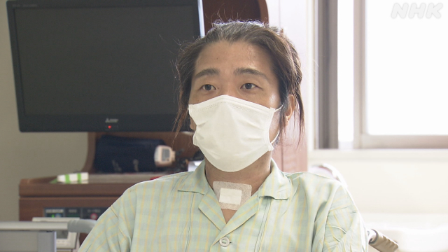
A man with severe respiratory failure.
I was treated in the ICU = intensive care unit for two months and miraculously recovered.
What was the difference between the life and death of men?
(Osaka base broadcasting station reporter Yuya Inagaki Daimu Shimizu)
I can't breathe through my torn lungs
Mr. Asayoshi Kubota, who lives in Osaka Prefecture, talked about his experience of fighting illness.
The infection was discovered immediately after the Golden Week holidays in May.
I was 45 years old.
In the midst of the fourth wave, it was when the medical system in Osaka was the most tight.
Mr. Kubota says he has a history of smoking but has no underlying illness.
In addition to a fever of 39 degrees, I felt abnormal taste and smell and was examined.
Mr. Kubota, who lived alone, continued to receive medical treatment at home, but his fever did not go down and his breathing gradually became difficult.
Mr. Kubota:
"The fever of 39 degrees continued, and my breathing became very difficult, so I called an ambulance because I was afraid. But I couldn't find the hospital, so I waited for hours while moaning in the ambulance. I have a memory. "
It was finally transported to a local general hospital.
It was nine days after the infection was discovered.
Although he was diagnosed with severe respiratory failure and was treated, his symptoms did not improve.
Two weeks later, I was forced to transfer to the Kansai Medical University Medical Center, where advanced treatment is possible.
Black cavity where the lungs should be
(Image: The lungs are crushed and a black cavity with nothing is expanding.)
This is a CT image of the lungs immediately after the transfer.
On CT, areas with tissue such as organs and blood vessels are usually white, and areas with nothing are black.
In Mr. Kubota's image, there was a black cavity in the chest where the lungs should be.
The virus destroyed the tissue of the lungs, punctured them, crushed the lungs, and made them about one-third the size of their original size.
It was as if the inflated balloon had a hole and the air inside had deflated.
In the intensive care unit undergoing surgery to close the hole in the lungs
Mr. Kubota
"I was told that one of the lungs was torn and there was a hole in it. Anyway, it's tough. Even if I inhale or inhale, oxygen doesn't come in, so I can't breathe like I'm drowning. I feel did "
two days after hospitalization, Kubota's had the surgery to close the hole in the lung, went into intensive care.
At this time, I put on an artificial heart-lung machine = ECMO and a respirator.
I needed to rest my lungs until the damage from the virus was healed.
Kubota's memory is cut off here.
For the next two months, my medical condition went up and down.
The doctor in charge of the treatment
"I was despairing that it was really useless at one point," said
Dr. Yasushi Nakamori of the Kansai Medical University Medical Center, who was in charge of treatment for Mr. Kubota.
The center has accepted more than 800 patients since the first wave of last spring, and Dr. Nakamori, the deputy hospital director, has played a central role in medical care.
Approximately three weeks after the surgery to close the hole in the lungs, Mr. Kubota's lungs, which had been recovering after the surgery, were in a bad condition again.
It was not the "virus" that broke the lungs, but the "immune runaway"
(Image: The tissue inside the lungs was destroyed and turned into a black cavity)
Unlike before surgery, this time there was a black cavity inside the lungs.
The inflammation had progressed into the lungs and even the internal tissues had been destroyed.
The cause was not a virus.
When infected with the new corona, the "immunity" that is supposed to work to eliminate the virus and protect one's body may run away and attack one's own body.
It is considered to be one of the causes of the aggravation, and it seems that this is also the cause of inflammation of Mr. Kubota's lungs.
The lung cavity was large, and it was difficult to perform surgery to close it this time.
Many of the ICU staff who saw the images said that they could only do a "lung transplant" to replace the entire lung.
Life or death The focus was on how to use medicine
Is it possible to suppress the runaway of immunity and regain the function of the lungs?
We will reach the first mountain, which divides life and death.
The focus was on the use of anti-inflammatory drugs such as "dexamethasone."
Anti-inflammatory drugs suppress inflammation by suppressing the action of runaway immunity.
In particular, severely ill patients have been reported to recover from severe pneumonia after administration of the drug.
On the other hand, reducing immune function with anti-inflammatory drugs creates another problem.
Since the immunity that protects the body does not work, it becomes easy to get infected with bacteria that are originally in human skin and the environment.
In fact, after using anti-inflammatory drugs, bacterial infections occur, the symptoms worsen, and deaths occur one after another.
(Image: Mr. Kubota receiving treatment)
An anti-inflammatory drug that can be called a double-edged sword.
It is necessary to make a heavy decision on which drug to use at what timing, which can directly affect the life or death of the patient.
Dr. Nakamori decided to focus on risk awareness in order to reduce the inflammation of the lungs of Mr. Kubota, who suffered serious damage.
On the other hand, we also took measures to control infections such as bacteria.
In addition to the administration of antibacterial drugs, we responded by increasing the frequency of replacing catheter tubes, which are likely to be a route of bacterial infection, from weekly to every three days.
As a result of continuing treatment while carefully observing the state of inflammation and the presence or absence of bacterial infection, Mr. Kubota's lungs gradually recovered.
The bacterial infection I was worried about was somehow suppressed, and pneumonia subsided in mid-July, a month and a half after admission.
Then, we will reach the peak of the second treatment.
It was how to remove the ECMO and ventilator.
The last challenge ECMO and how to remove the ventilator
(Image: Heart-lung machine = ECMO)
Actually, Mr. Kubota has removed the ECMO twice by this time.
However, in each case, the condition deteriorated immediately afterwards, and he repeatedly put it on again.
From this process, Dr. Nakamori thought that the ventilator used in combination had a part of the cause.
ECMO is a treatment that temporarily substitutes the function of the lungs by sending oxygen directly to the blood taken from the patient and returning the blood to the body.
There is a risk of blood clots on the way through the ECMO device, so it should be removed immediately after lung recovery.
(Image: Mr. Kubota wearing a ventilator and ECMO)
On the other hand, the ventilator also helps breathing by sending oxygen to the lungs, but if ECMO is removed first, the amount supplemented by ECMO will decrease. Instead, the amount delivered through the ventilator will increase.
Dr. Nakamori thought that Mr. Kubota's lungs could not withstand the sudden change in the burden.
So, this time, which was the third time, I decided to remove the ventilator first.
Similarly, reports of cases of recovery by removing the ventilator first also helped the decision.
Ventilator disengagement and return to consciousness The season is from spring to summer
Mr. Kubota regained consciousness after the ventilator was removed.
This is a picture taken in the intensive care unit with the ECMO tube connected to the neck.
The season was from spring to summer.
Dr. Nakamori's decision was successful, and Mr. Kubota was able to return to the general ward two weeks later.
Kubota's life and death was divided by the attitude of the medical team, who continued to find solutions even when they were forced into a difficult situation.
Dr. Yasushi Nakamori, Kansai Medical University Medical Center
"I really felt the limit of treatment when I had a cavity in my lungs and couldn't operate. I thought it was a last resort, but I used an anti-inflammatory drug. The process of recovery can only be described as a miracle. I feel that Mr. Kubota once again taught me that if I treat him without giving up, he will be cured. "
Mr. Kubota was discharged from the hospital in late August, and then. I continued to rehabilitate while going to the hospital.
Calls from medical staff helped in a fierce fight against illness
(Image: Comparison of CT images of the lungs) The lung tissue was restored immediately before discharge
.) The lung tissue of Mr. Kubota, who had a hole immediately after admission and was seriously damaged as the size shrank.
After that, by closing the hole in the lungs by surgery and repairing it over time, it was gradually regaining its original function.
The CT image taken just before discharge clearly showed the lung tissue where it should be.
There is one more thing that supported Mr. Kubota's life in the fierce fight against illness over the two months.
In the turbid consciousness, Mr. Kubota's memory was a call from the ICU staff.
Mr. Kubota
"I faintly remember that the nurse next to the bed often called on and encouraged Mr. Kubota and Mr. Kubota. It was a great support to me and I am really grateful. I think I was in a dying state, but I want to do my best so that I can live my original life without being impatient. But I want you to know that there are patients who can be saved and hope for treatment. "