
As you read this article, a ray of light crosses your cornea, traveling through your pupil and lens until it reaches your retina;
Where there are light-sensing cells - such as rods and cones - which in turn pass this visual information to the brain after translating it into electrical signals in the optic nerve, and then vision occurs.
new lenses
However, a recent study - published in the journal Science Advances on the second of this March - has added a new lens to this process.
The study indicated that mitochondria - which are cellular organelles responsible for producing energy in the cell - act as tiny lenses to help deliver photons of light to neurons.
According to the press release published by the US National Institutes of Health, Dr. Wei Lee - neurologist and senior author of the study - says, "We were amazed by this amazing phenomenon that shows a dual role for mitochondria; in addition to their well-established metabolic function that produces energy, It plays a role in the vision process.”
Mitochondria are amazing microorganisms;
As it undertakes the production of energy, and then enables the cell to complete its functions.
And if you remember its shape from biology courses, you definitely know that it is not that structure that might seem effective in directing light.
In this study, the researchers used the striped ground squirrel as an ideal model to study this phenomenon;
These non-nocturnal animals have many cone cells to distinguish colors.
They also don't have many rods, at least compared to other mammals, to see in the dark, which allowed the researchers to explore an isolated layer of these cone cells containing live mitochondria.
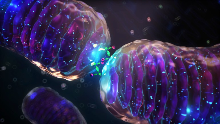
Mitochondria are amazing tiny organelles involved in the production of energy in the cell (Getty Images)
special focus
To reach these results, the retina was dissected into 3 parts, and then all layers except for the light-sensitive cells (photoreceptors) were removed to be examined under a microscope.
The researchers highlighted these photoreceptors, which contain live mitochondria, enabling them to explore their new role as a microlens.
This seemed strange to the researchers, as they noticed that mitochondria are located in the outer light-sensing part of the retinal cone cells;
Hence, this means that photons of light can directly hit the mitochondria, which can cause the light to be scattered in different directions or even absorbed.
This will eventually prevent it from reaching the nerve cells.
These complex, lipid-rich organelles will also influence the passage of light, the researchers say.
But although light can be scattered or absorbed by these organelles, the researchers showed - using microscopic imaging techniques and simulations - that mitochondria focus light to fall on the outside of the retinal cone cells.
Hence, this tight positioning of mitochondria helps direct and focus light on retinal cells.
The tight placement of mitochondria helps direct and focus light on retinal cells (Getty Images)
Stiles-Crawford property
Although mammalian cone cells are very similar, we cannot be certain that this happens in human cells either.
Therefore, we need more research.
According to the report, published by Science Alert, John Paul - the study's first author - states that this lens-like function may also explain a phenomenon known as the Stiles-Crawford property.
The Stiles-Crawford property is a phenomenon specific to cone receptors, which states that light entering the center of the pupil triggers a response in the cone cells more than light that passes near the edge of the pupil.
The team discovered - with experimental evidence and through simulation models - that the interaction of mitochondria with light matches the property of "Stiles-Crawford", which means that mitochondria may be the cause of this phenomenon.