
Share
02 April 2020 The famous 'crown' can also be seen in the first black and white portrait of the SarsCoV2 virus isolated in Lombardy at the Sacco Hospital in Milan: electron microscopic images show the viral particles attached to cell membranes and their typical crown of surface glycoproteins. The announcement in a note from the State University of Milan.The coronavirus had been isolated at the Laboratory of Infectious Diseases by the group coordinated by Massimo Galli and Gianguglielmo Zehender, in collaboration with the Pathological Anatomy directed by Manuela Nebuloni of the Department of Biomedical and Clinical Sciences Luigi Sacco. The isolations were obtained by researchers Alessia Lai, Annalisa Bergna, Arianna Gabrieli and Maciej Tarkowski, while Antonella Tosoni and Beatrice Marchini made the observations under the electron microscope and produced the beautiful images.
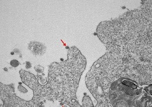
In the first, at a magnification of 30000X, the viral particles of SarsCov2 (indicated by arrows) are observed, attached to the membranes on the surface and inside cells used for isolation. The combination of two other images at different magnifications (50000X and 140000X) shows the viral particles with the typical ultrastructure characterized by the crown of surface glycoproteins.